A recent preprint by Dr. Anil Wadhwani and Dr. David Goldberg presents machine learning models capturing brain p-tau in different pathological contexts.
Alzheimer’s disease is marked by buildups of proteins within and around brain cells that can impede function and eventually lead to cell death. Two proteins, Amyloid-beta (AB) and Tau, can each form aggregates that are difficult to measure, predict, and precisely map to disease outcomes.
Tau becomes a risk for disease when a phosphate group is added, turning it into phosphorylated tau (p-tau), allowing it to form neurofibrillary tangles (NFTs) inside cells. The combination of AB plaques (outside of cells) and NFTs is typical of Alzheimer’s disease (AD), but NFTs can independently lead to a related condition called Primary Age Related Tauopathy (PART). PART may lead to some cognitive issues but has a distinct molecular profile from Alzheimer’s disease.
PART and AD can be differentiated by the spread of p-tau through the brain’s different regions. In PART, p-tau stays in regions like the hippocampus whereas in AD it spreads to neighboring cortex regions.
Understanding why p-tau stays in the hippocampus in PART but spreads to neocortical regions in AD can inform the differences in trajectories of brain aging.
Dr. Anil Wadhwani and Dr. David Goldberg are joint lead authors of a recent pre-print investigating this question using DNA methylation profiles across brain regions.
When a C is followed by a G in the human genome (often called a CpG site), it can be methylated as a form of epigenetic regulation. This modification can increase or decrease nearby gene expression but is highly context dependent. As the genetic architectures of AD and PART strongly overlap, DNA methylation patterns may help distinguish these related conditions.
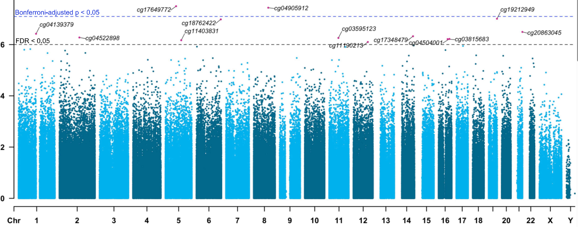
The authors identified 13 CpG sites where methylation was newly associated with hippocampal p-tau. Some of these sites were also associated with expression changes in up to 198 genes. These genes (shown in Figure 1 below) are related to processes like ion transport, synaptic transmission, and neuron architecture; processes relevant to Alzheimer’s disease.
 Figure 1: Figure 1c from the MedRxiv manuscript. One CpG site, labeled as cg04522898, is positively (red lines) correlated with known gene networks.
Previous work has identified DNAm and other epigenetic markers associated with aging, allowing the development of epigenetic clocks to distinguish chronological aging from biological aging. To build on this work, the authors use brain region-specific DNAm signals to monitor the spread and severity of p-Tau in the brain, and how these relate to Alzheimer’s Disease.
The new epigenetic clocks the authors developed are called TauSeverity and TauSpread. These clocks are machine learning models trained on CpG methylation levels to predict hippocampal p-tau and the spread of p-tau from the hippocampus to cortex regions, respectively. These models were trained using a special type of linear regression model called an Elastic Net (EN) and were trained on 470 CpG sites each with only 8 CpG sites in common between them.
Although developed to predict severity of p-tau in the hippocampus and its spread to the cortex, the TauSeverity and TauSpread models could effectively distinguish between individuals with or without cognitive impairment. Beyond this, the authors developed a support vector machine (SVM) model called ResilienceDetector that uses 1,214 CpG sites to distinguish PART and AD cases.
When studying Alzheimer’s disease, ‘resilience’ refers to showing minimal cognitive deficits despite pathological markers of disease. In this context, individuals with comparable levels of p-tau showed significant variability in cognitive function.
The ResilienceDetector model could successful separate most cases and revealed that the groups labelled as ‘high resilience’ and ‘low resilience’ had comparable p-tau levels and TauSeverity/TauSpread scores. This may show that ResilienceDetector is picking up on a different epigenetic signature.
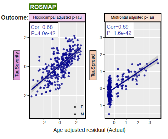
Figure 2. Figure 2a from the MedRxiv manuscript. TauSeverity and TauSpread models predict hippocampal and midfrontal p-tau levels, after adjusting for age, in the ROSMAP cohort. Cor indicates Pearson’s correlation coefficient.
These DNAm models enable investigation into molecular mechanisms that distinguish PART from AD, and in the future, may be applied more broadly to other tissue types, like blood.
The TauSpread and TauSeverity models will be available upon final publication of this work. For more details, please see the manuscript available on MedRxiv https://doi.org/10.1101/2024.11.07.24316933.
Featured authors
Anil Wadhwani, MD, PhD
Dr. Wadhwani is an Instructor in the Department of Neurology and a post-doctoral fellow in the Penn Bioinformatics in Neurodegenerative Disease (BiND) lab, led by Dr. Corey McMillan.
David Goldberg, PhD
Dr. Goldberg is a recent graduate of the Neuroscience Graduate Group who completed his doctoral thesis in Dr. Wanding Zhou’s lab in the Children’s Hospital of Philadelphia.